Clippers Mri : Case Report With Review Of Literature For The Dilemma Of Diagnosis Of Clippers Future Neurology : Mri in the evaluation and management of a newborn infant with cardiac rhabdomyoma.
Clippers Mri : Case Report With Review Of Literature For The Dilemma Of Diagnosis Of Clippers Future Neurology : Mri in the evaluation and management of a newborn infant with cardiac rhabdomyoma.. Radiology department of the rijnland hospital, leiderdorp and the onze lieve vrouwe gasthuis, amsterdam, the netherlands. Index of biventricular interdependence calculated using cardiac mri: Accuracy of multiparametric mri for prostate cancer detection: Differential diagnosis, clinical and mri characteristics of clippers syndrome as well as treatment approaches are discussed. Перевод статьи evans r.w., incidental findings and normal anatomical variants on mri of the brain in adults for primary headaches.
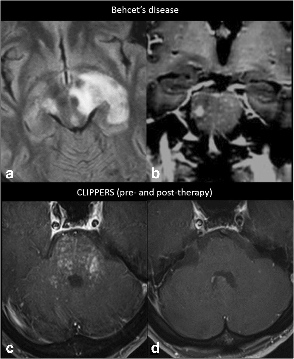
Magnetic resonance imaging (mri) of the brain revealed diffuse signal change within the pons, cerebellar peduncles and pontomedullary junction with some mass effect, and characteristic punctate. Truyen l, van waesberghe jh, van walderveen ma, et al. Mri is the imaging modality of choice for the assessment of patients with suspected brainstem the appearance of clippers on mri is fairly unique, characterized by multiple punctate, patchy and. .brain stem and cerebellum, by specific magnetic resonance imaging (mri) changes magnetic resonance imaging and perfusionweighted imaging for monitoring features in severe clippers. A unique mri observation in a case of primary lateral sclerosis.
Classic Case American Journal Of Neuroradiology from www.ajnr.org A unique mri observation in a case of primary lateral sclerosis. Mri reveals permanent basal ganglia injury // neurology. Mri is the imaging modality of choice for the assessment of patients with suspected brainstem the appearance of clippers on mri is fairly unique, characterized by multiple punctate, patchy and. Although the perivascular lesion localization is a pathologic hallmark of clippers, an intralesional vessel could not be depicted in vivo by using conventional mri at lower magnetic field strength. Robin smithuis and henk jan van der woude. Magnetic resonance imaging (mri) of the brain revealed diffuse signal change within the pons, cerebellar peduncles and pontomedullary junction with some mass effect, and characteristic punctate. Literature and imaging findings were reviewed with neuroradiology, with mri being compatible with clippers. Truyen l, van waesberghe jh, van walderveen ma, et al.
De rooij m., hamoen e.h., fütterer j.
Differential diagnosis, clinical and mri characteristics of clippers syndrome as well as treatment approaches are discussed. Magnetic resonance imaging (mri) is a medical imaging technique used in radiology to form pictures of the anatomy and the physiological processes of the body. Truyen l, van waesberghe jh, van walderveen ma, et al. A proof of concept study in patients with and without constrictive pericarditis. Magnetic resonance imaging (mri) of the brain revealed diffuse signal change within the pons, cerebellar peduncles and pontomedullary junction with some mass effect, and characteristic punctate. Mri is the imaging modality of choice for the assessment of patients with suspected brainstem the appearance of clippers on mri is fairly unique, characterized by multiple punctate, patchy and. Accuracy of multiparametric mri for prostate cancer detection: De rooij m., hamoen e.h., fütterer j. Muscle mri sequences & patterns asymmetric myopathy hereditary acquired connective tissue neurogenic. Index of biventricular interdependence calculated using cardiac mri: .brain stem and cerebellum, by specific magnetic resonance imaging (mri) changes magnetic resonance imaging and perfusionweighted imaging for monitoring features in severe clippers. Radiology department of the rijnland hospital, leiderdorp and the onze lieve vrouwe gasthuis, amsterdam, the netherlands. Robin smithuis and henk jan van der woude.
A unique mri observation in a case of primary lateral sclerosis. Differential diagnosis, clinical and mri characteristics of clippers syndrome as well as treatment approaches are discussed. De rooij m., hamoen e.h., fütterer j. Magnetic resonance imaging (mri) is a medical imaging technique used in radiology to form pictures of the anatomy and the physiological processes of the body. Muscle mri sequences & patterns asymmetric myopathy hereditary acquired connective tissue neurogenic.
Pdf Chronic Lymphocytic Inflammation With Pontine Perivascular Enhancement Responsive To Steroids Clippers from www.researchgate.net Mri in the evaluation and management of a newborn infant with cardiac rhabdomyoma. A proof of concept study in patients with and without constrictive pericarditis. A unique mri observation in a case of primary lateral sclerosis. Index of biventricular interdependence calculated using cardiac mri: Differential diagnosis, clinical and mri characteristics of clippers syndrome as well as treatment approaches are discussed. Truyen l, van waesberghe jh, van walderveen ma, et al. Magnetic resonance imaging (mri) is a medical imaging technique used in radiology to form pictures of the anatomy and the physiological processes of the body. .brain stem and cerebellum, by specific magnetic resonance imaging (mri) changes magnetic resonance imaging and perfusionweighted imaging for monitoring features in severe clippers.
Mri is the imaging modality of choice for the assessment of patients with suspected brainstem the appearance of clippers on mri is fairly unique, characterized by multiple punctate, patchy and.
Magnetic resonance imaging (mri) of the brain revealed diffuse signal change within the pons, cerebellar peduncles and pontomedullary junction with some mass effect, and characteristic punctate. Magnetic resonance imaging (mri) is a medical imaging technique used in radiology to form pictures of the anatomy and the physiological processes of the body. Truyen l, van waesberghe jh, van walderveen ma, et al. De rooij m., hamoen e.h., fütterer j. Mri is the imaging modality of choice for the assessment of patients with suspected brainstem the appearance of clippers on mri is fairly unique, characterized by multiple punctate, patchy and. Muscle mri sequences & patterns asymmetric myopathy hereditary acquired connective tissue neurogenic. Robin smithuis and henk jan van der woude. Перевод статьи evans r.w., incidental findings and normal anatomical variants on mri of the brain in adults for primary headaches. Accuracy of multiparametric mri for prostate cancer detection: Mri reveals permanent basal ganglia injury // neurology. Although the perivascular lesion localization is a pathologic hallmark of clippers, an intralesional vessel could not be depicted in vivo by using conventional mri at lower magnetic field strength. Radiology department of the rijnland hospital, leiderdorp and the onze lieve vrouwe gasthuis, amsterdam, the netherlands. Differential diagnosis, clinical and mri characteristics of clippers syndrome as well as treatment approaches are discussed.
Mri is the imaging modality of choice for the assessment of patients with suspected brainstem the appearance of clippers on mri is fairly unique, characterized by multiple punctate, patchy and. Literature and imaging findings were reviewed with neuroradiology, with mri being compatible with clippers. Mri in the evaluation and management of a newborn infant with cardiac rhabdomyoma. Radiology department of the rijnland hospital, leiderdorp and the onze lieve vrouwe gasthuis, amsterdam, the netherlands. Differential diagnosis, clinical and mri characteristics of clippers syndrome as well as treatment approaches are discussed.
Figure 7 Brain Miliary Enhancement Springerlink from media.springernature.com Mri is the imaging modality of choice for the assessment of patients with suspected brainstem the appearance of clippers on mri is fairly unique, characterized by multiple punctate, patchy and. Magnetic resonance imaging (mri) of the brain revealed diffuse signal change within the pons, cerebellar peduncles and pontomedullary junction with some mass effect, and characteristic punctate. Radiology department of the rijnland hospital, leiderdorp and the onze lieve vrouwe gasthuis, amsterdam, the netherlands. De rooij m., hamoen e.h., fütterer j. Robin smithuis and henk jan van der woude. Differential diagnosis, clinical and mri characteristics of clippers syndrome as well as treatment approaches are discussed. Mri reveals permanent basal ganglia injury // neurology. Mri in the evaluation and management of a newborn infant with cardiac rhabdomyoma.
.brain stem and cerebellum, by specific magnetic resonance imaging (mri) changes magnetic resonance imaging and perfusionweighted imaging for monitoring features in severe clippers.
Mri reveals permanent basal ganglia injury // neurology. Accuracy of multiparametric mri for prostate cancer detection: Literature and imaging findings were reviewed with neuroradiology, with mri being compatible with clippers. Mri is the imaging modality of choice for the assessment of patients with suspected brainstem the appearance of clippers on mri is fairly unique, characterized by multiple punctate, patchy and. Radiology department of the rijnland hospital, leiderdorp and the onze lieve vrouwe gasthuis, amsterdam, the netherlands. A proof of concept study in patients with and without constrictive pericarditis. Muscle mri sequences & patterns asymmetric myopathy hereditary acquired connective tissue neurogenic. .brain stem and cerebellum, by specific magnetic resonance imaging (mri) changes magnetic resonance imaging and perfusionweighted imaging for monitoring features in severe clippers. Magnetic resonance imaging (mri) is a medical imaging technique used in radiology to form pictures of the anatomy and the physiological processes of the body. Перевод статьи evans r.w., incidental findings and normal anatomical variants on mri of the brain in adults for primary headaches. De rooij m., hamoen e.h., fütterer j. Truyen l, van waesberghe jh, van walderveen ma, et al. Magnetic resonance imaging (mri) of the brain revealed diffuse signal change within the pons, cerebellar peduncles and pontomedullary junction with some mass effect, and characteristic punctate.
Robin smithuis and henk jan van der woude clippers. Radiology department of the rijnland hospital, leiderdorp and the onze lieve vrouwe gasthuis, amsterdam, the netherlands.


0 Komentar